
The ultrasound performed at 5 weeks of amenorrhea (WA) is one of the earliest examinations in pregnancy monitoring. At this stage, the central question is what the image actually reveals: which structures are measurable, which remain invisible, and most importantly, what conclusions can be drawn without error?
β-hCG Threshold and Visibility of the Gestational Sac on Ultrasound
The visibility of the gestational sac on transvaginal ultrasound directly depends on the level of β-hCG. Before 5 WA, the sac becomes detectable when the level of β-hCG is greater than or equal to 1,000 IU. Below this threshold, the examination may show nothing, without indicating an anomaly.
At 5 WA, the gestational sac measures a few millimeters. It is this measurement of the sac’s diameter that allows for estimating gestational age at this very early stage, before the embryo is measurable. The information provided by the ultrasound at 5 WA and gestational sac remains limited but significant: it confirms the intrauterine location of the pregnancy and rules out an ectopic pregnancy.
On the other hand, a high level of β-hCG without a visible sac on ultrasound is a warning signal that justifies close monitoring.
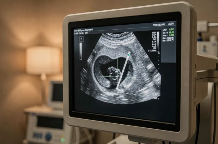
Visible and Non-Visible Structures at 5 WA: Comparative Table
The most common confusion at this stage is expecting information from the ultrasound that it cannot yet provide. The table below summarizes what is observable and what is not.
| Structure | Visible at 5 WA? | Details |
|---|---|---|
| Gestational sac | Yes (if β-hCG sufficient) | Diameter of a few mm, surrounded by the trophoblastic ring |
| Yolk sac | Sometimes | May appear at the end of 5 WA, not systematic |
| Embryo (embryonic echoes) | Rarely | Visible when the sac reaches about 10 mm |
| Cardiac activity | No | Generally detectable from 6-7 WA |
| Primitive brain vesicles | No | Appear around 8 WA |
The yolk sac, when spotted, constitutes the first sign of a confirmed intrauterine pregnancy. Its presence inside the sac is more reassuring than the sac alone.
Why Not Conclude a Clear Egg from a Single Ultrasound at 5 WA
A empty gestational sac at 5 WA often causes immediate concern. The absence of a visible embryo sometimes leads to a premature diagnosis of a stopped pregnancy or clear egg. Recent international recommendations are categorical on this point.
The Society of Radiologists in Ultrasound (SRU), ACOG, and NICE recommend never diagnosing a stopped pregnancy based on a single ultrasound performed before 7 WA. These scientific societies systematically advocate for a follow-up ultrasound at least 7 to 14 days after the first.
This waiting period allows for verifying two elements:
- The growth of the gestational sac, which should increase steadily between the two examinations
- The possible appearance of an embryo with cardiac activity, expected around 6-7 WA when the embryo reaches a few millimeters
- The concordance between the evolution of the β-hCG level and the ultrasound data, as a stagnant or decreasing level indicates a non-evolving pregnancy
In practical terms, a empty sac at 5 WA is not a diagnosis, it is an observation to be monitored. The area between 5 and 7 WA is considered “probabilistic”: the data is insufficient to make a definitive conclusion either way.

Measurement of the Gestational Sac and Early Pregnancy Dating
At 5 WA, dating relies exclusively on the average diameter of the gestational sac. The embryo is not yet measurable in most cases, so the crown-rump length (CRL), which will become the reference for dating in the first trimester, cannot be used.
The measurement of the gestational sac remains less precise than the CRL for dating a pregnancy. A margin of error of several days is common. This is why the definitive dating will be recalculated during the first trimester ultrasound, between 11 and 14 WA, when the CRL of the embryo can be reliably measured.
At 7 WA, the embryo measures about 10 mm and shows cardiac activity. The yolk sac is then clearly individualized. At 8 WA, the embryo reaches about 17 mm, and several structures (primitive brain vesicles, stomach) become identifiable.
Correlation Between β-hCG Levels and Early Ultrasound
The link between β-hCG and ultrasound works both ways. A level that doubles regularly every 48 hours is reassuring, even if the ultrasound shows only an empty sac. A stagnant level with a small sac points towards an unfavorable outcome.
The relationship is not linear. Two normal evolving pregnancies can present very different β-hCG levels at the same date. It is the dynamic progression of the level that matters, not its absolute value.
- A level below the ultrasound detection threshold (less than 1,000 IU) explains an “empty” examination without indicating an abnormal pregnancy
- A high level without a visible intrauterine sac necessitates searching for an ectopic pregnancy
- A level that progresses normally with a visible sac but no embryo at 5 WA justifies a simple follow-up at 7-14 days
The interpretation of the ultrasound at 5 WA is never done in isolation. It only makes sense when coupled with the β-hCG measurement and, in most cases, a follow-up examination. A single image is not enough to confirm or rule out the viability of a pregnancy at this early stage.